
Introduction
Posterior infarction accompanies 15-20% of STEMIs, usually occurring in the context of an inferior or lateral infarction
- Isolated posterior MI is less common (3-11%)
- Posterior extension of an inferior or lateral infarct implies a much larger area of myocardial damage, with an increased risk of left ventricular dysfunction and death
Etiology
Usually caused by posterior descending artery (PDA) occlusion.
Approximately 70% of the population is “right-dominant” (RCA supplies the PDA), while 10% are “left-dominant (the LCx supplies the PDA), and 20% are “co-dominant” (both the RCA and LCx jointly supply the PDA).
Pathophysiology
Rupture of a coronary artery plaque, thrombosis, and blockage of the downstream perfusion leading to myocardial ischemia and necrosis.
ECG manifestation
Posterior myocardium is not directly visualised by 12-lead ECG, given the placement of anteroseptal leads V1-V3, they are indirectly examining the posterior wall too
- Horizontal ST depressions V1-V3
- Dominant R wave (R/S ratio > 1) in V2-V3
- Large/upright broad R waves (>30ms)
- Upright T waves V1-V3
Posterior leads: Posterior MI is confirmed by the presence of ST elevation in the posterior leads (V7-9). The degree of ST elevation seen in V7-9 is typically modest – only 0.5 mm of ST elevation is required to make the diagnosis of posterior MI!
Picture 1 Posterior leads
Lead V7: posterior axillary line
Lead V8: midscapular
Lead V9: paraspinal

https://litfl.com/posterior-myocardial-infarction-ecg-library/
ECG 1 STEMI of posterior wall (ST depressions and upright T waves in V1-V3)
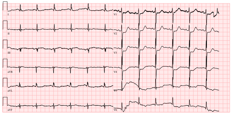
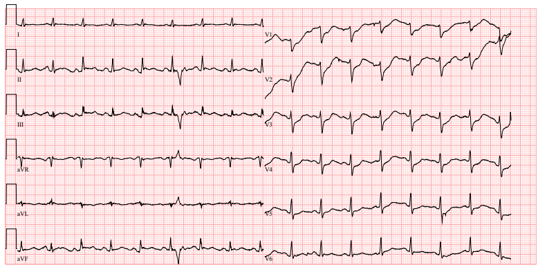
ECG 2 STEMI of posterior and inferior wall (ST depressions in V1-V3, STE in II, III, aVF)
Management
- coronary angiography/PCI
- UFH
- ASA
- P2Y12 inhibitors
- nitrates
- analgesia
- 02
References
- Brady W, Erling B, Pollack M, et al. Electrocardiographic manifestations: acute posterior wall myocardial infarction. J Emerg Med 2001;20:391-401
- Lizzo JM, Chowdhury YS. Posterior Myocardial Infarction. 2020 Nov 29. In: StatPearls [Internet]. Treasure Island (FL): StatPearls Publishing; 2020 Jan–. PMID: 31985961.
- http://www.emdocs.net/ecg-pointers-posterior-mi/
- https://litfl.com/posterior-myocardial-infarction-ecg-library/

Introduction
Posterior infarction accompanies 15-20% of STEMIs, usually occurring in the context of an inferior or lateral infarction
- Isolated posterior MI is less common (3-11%)
- Posterior extension of an inferior or lateral infarct implies a much larger area of myocardial damage, with an increased risk of left ventricular dysfunction and death
Etiology
Usually caused by posterior descending artery (PDA) occlusion.
Approximately 70% of the population is “right-dominant” (RCA supplies the PDA), while 10% are “left-dominant (the LCx supplies the PDA), and 20% are “co-dominant” (both the RCA and LCx jointly supply the PDA).
Pathophysiology
Rupture of a coronary artery plaque, thrombosis, and blockage of the downstream perfusion leading to myocardial ischemia and necrosis.
ECG manifestation
Posterior myocardium is not directly visualised by 12-lead ECG, given the placement of anteroseptal leads V1-V3, they are indirectly examining the posterior wall too
- Horizontal ST depressions V1-V3
- Dominant R wave (R/S ratio > 1) in V2-V3
- Large/upright broad R waves (>30ms)
- Upright T waves V1-V3
Posterior leads: Posterior MI is confirmed by the presence of ST elevation in the posterior leads (V7-9). The degree of ST elevation seen in V7-9 is typically modest – only 0.5 mm of ST elevation is required to make the diagnosis of posterior MI!
Picture 1 Posterior leads
Lead V7: posterior axillary line
Lead V8: midscapular
Lead V9: paraspinal
https://litfl.com/posterior-myocardial-infarction-ecg-library/
ECG 1 STEMI of posterior wall (ST depressions and upright T waves in V1-V3)
ECG 2 STEMI of posterior and inferior wall (ST depressions in V1-V3, STE in II, III, aVF)
Management
- coronary angiography/PCI
- UFH
- ASA
- P2Y12 inhibitors
- nitrates
- analgesia
- 02
References
- Brady W, Erling B, Pollack M, et al. Electrocardiographic manifestations: acute posterior wall myocardial infarction. J Emerg Med 2001;20:391-401
- Lizzo JM, Chowdhury YS. Posterior Myocardial Infarction. 2020 Nov 29. In: StatPearls [Internet]. Treasure Island (FL): StatPearls Publishing; 2020 Jan–. PMID: 31985961.
- http://www.emdocs.net/ecg-pointers-posterior-mi/
- https://litfl.com/posterior-myocardial-infarction-ecg-library/


